Category Archives: Ear Presentation

Embryology

Development of the ear begins during the first few weeks of pregnancy. At this time the developing fetus looks like a FISH with gills.

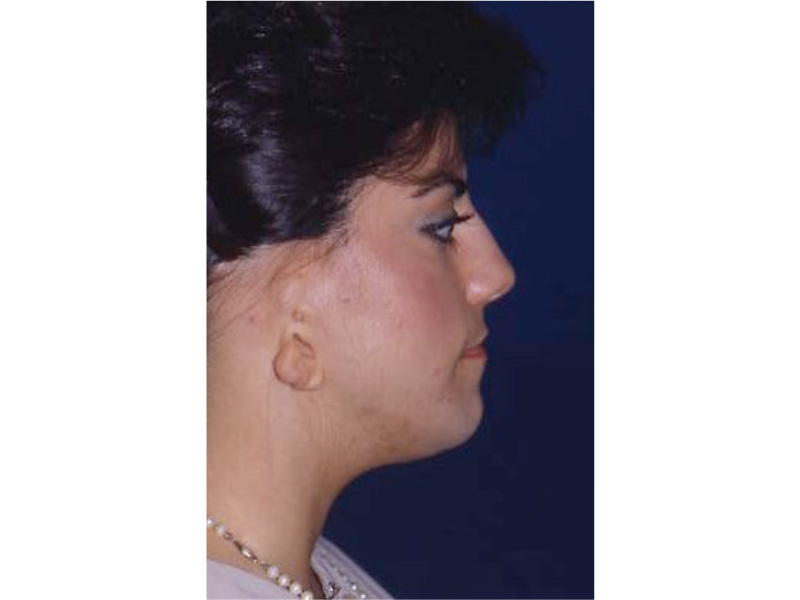
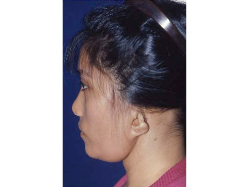
As the shape of the auricle forms, the developing ear moves (migrates) from under the chin to its proper position on the skull. These two patients have auricles that failed to completely move from under the chin to the skull.
This slide shows the proper position of the ear. The auricle should attain its adult size by age 8. The auricle should lie between the eyebrow and the bottom of the nose. It should not lie straight, but be parallel to the bride of the now. It should also lie approximately 6 cm (the average height of an adult ear) from the eye.
info_on_the_ear_0048_1embryology
info_on_the_ear_0049_1embryology

As  the ear is developing in the first few weeks  of gestation its shape not only changes to that  of a normal ear, but it moves from under the chin to it proper position on the skull.  These two patient’s developing ears (auricles) failed to adequately move to their proper adult position.
info_on_the_ear_0050_1embryology

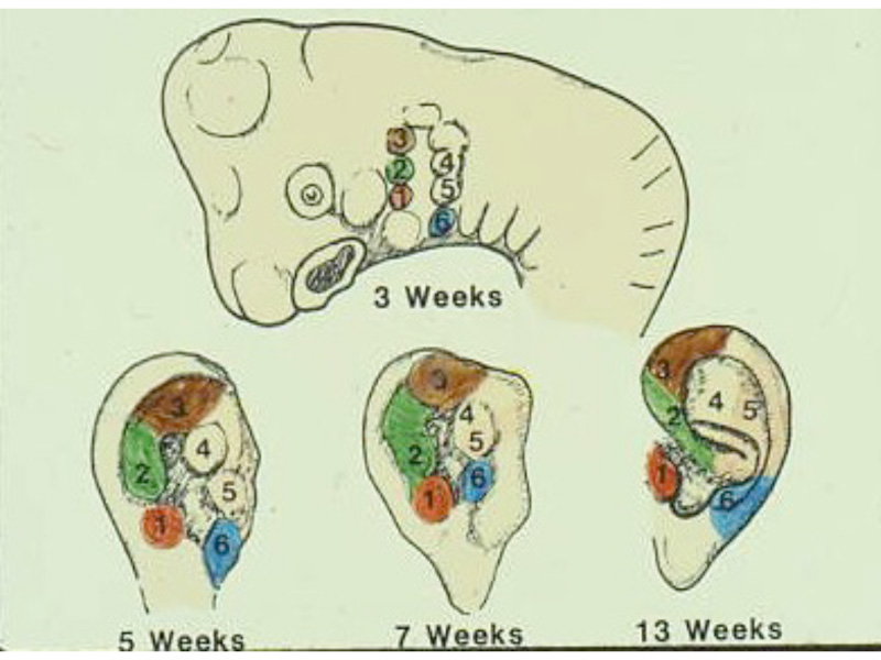
These diagrams picture the developing ear (auricle) from the first three weeks of pregnancy through the end of the third month of pregnancy.  Initially the ear looks like “gills” on a fish.  By the end of the third month of pregnancy, the ear has attained its adult shape.

Concomitant developing organ systems

Other organ systems are also developing at the same time as the ear. Therefore the doctor must evaluate a Microtia child’s kidneys, heart, neck, and middle ear that may also be abnormally affected.

Etiology

Situations that may cause a child to have Microtia are

  1. Mother having fevers during the first few months of pregnancy.
  2. Any genetic predisposition (family history of others with ear anomalies).
  3. Mother having exposure to medications/environments that may affect development of the ear.
    • Accutane
    • Exposure to excessive radiation.
    • Thalidomide

Incidence

Microtia occurs in 1:7,000 children.
30% of affected children have Microtia in BOTH ears.
30% of affected children have OTHER congenital anomalies that must be searched for.

Evaluation of susceptible organ systems

Several tests may be necessary to find other anomalies:

  1. Ultrasound of the kidneys.
  2. Heart beats.
  3. X rays of the neck
  4. Hearing testing

Treacher Collins Syndrome

This syndrome has several other issues including Microtia and hearing loss.info_on_the_ear_0046_Treacher Collins

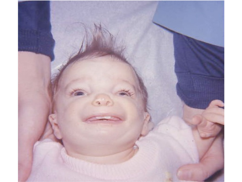
Patients with Treacher Collins Syndrome have other abnormalities of their face.  Their ears are ”low set”, and their cheek bones have not properly developed.


Goldenhar’s Syndrome

These patients often also have problems with their kidneys.

info_on_the_ear_0045_Goldenhar’s Syndrome

This syndrome represents other developmental problems that can occur at the same time the outer ear (auricle) is developing.  The facial nerve  (nerve to the face) has not developed causing a paralysis of this patient’s right side of  his face.  The ears are also “low set” and mal-developed. Also there are often problems with the kidneys with these patients.

Mandible and TMJ

Often one side of the face and jaw are smaller than the normal opposite side. This phenomenon is called “hemi-facial microsomia”.

info_on_the_ear_0044_Mandible

Anatomy

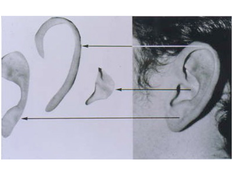
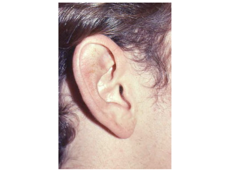
The auricle is not round, triangular, or square, but OVAL shaped. It is not flat, but is three dimensional consisting of THREE layers (concha, scapha, and helix).

info_on_the_ear_0042_Anatomy info_on_the_ear_0043_Anatomy

Evaluation

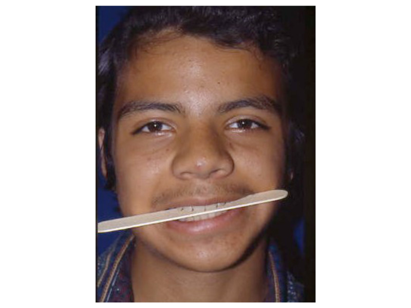
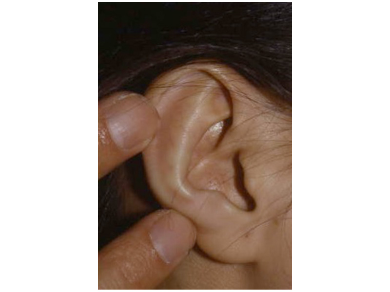
Using ones fingers many anomalies can be easily seen.

info_on_the_ear_0041_Evaluation